10 Tips for Choosing Mandibular Reconstruction Plates

Time:2026-10-27 Author:Sophia
0%

Selecting the right Mandibular Reconstruction Plates requires more than comparing sizes or prices. The decision affects facial symmetry, chewing function, speech, and long-term stability. Surgeons must assess bone loss, soft-tissue condition, occlusion, and the patient’s overall health before choosing a system.

Small details matter. A plate should follow the mandibular contour without creating sharp pressure points. Screw length must support fixation while avoiding tooth roots, the inferior alveolar canal, and nearby anatomical structures. Preoperative CT imaging can reveal defects that ordinary radiographs may miss. In complex cases, virtual planning and patient-specific solutions may improve accuracy, although they do not replace surgical judgment.

There is no universal plate for every reconstruction. Material strength, fatigue resistance, corrosion performance, and compatibility with available instruments deserve careful review. Experienced teams also consider flap design, bone quality, postoperative loading, and the risk of infection or hardware exposure. Manufacturer data and peer-reviewed evidence can guide these choices, but clinical experience remains essential.

Some decisions are less clear than they appear. Imaging may be incomplete. Intraoperative anatomy can differ from the surgical plan. A technically strong plate may still fail if contouring, fixation, or soft-tissue coverage is inadequate. This guide presents ten practical tips for evaluating Mandibular Reconstruction Plates with greater consistency. It also encourages thoughtful reassessment, because reliable reconstruction depends on both precise planning and careful adaptation during surgery.
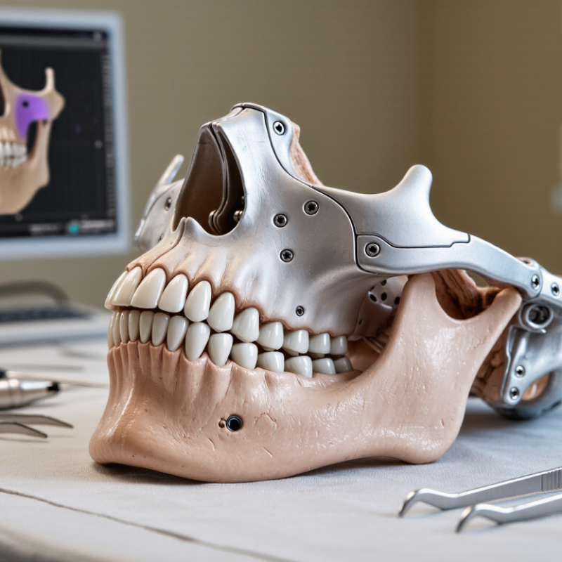
10 Tips for Choosing Mandibular Reconstruction Plates

Understanding the Role of Mandibular Reconstruction Plates

10 Tips for Choosing Mandibular Reconstruction Plates

Understanding the Role of Mandibular Reconstruction Plates

Mandibular reconstruction plates do more than bridge a bony defect. They maintain facial contour, stabilize occlusion, and protect healing tissues. GLOBOCAN 2022 reported 389,846 new lip and oral cavity cancer cases worldwide. This burden makes dependable reconstruction planning clinically important. A plate should match defect length, bone quality, load direction, and soft-tissue coverage. Surgeons also assess screw purchase near the mandibular angle, condyle, and symphysis. Small positioning errors can change chewing mechanics.

Published systematic reviews commonly report plate exposure, infection, or loosening rates between 10% and 30%, depending on patient selection and reconstruction method. These figures are not universal. Smoking, radiation therapy, poor nutrition, and thin mucosa can increase risk. The AO Foundation’s surgical guidance emphasizes accurate contouring, stable fixation, and careful soft-tissue management. Imaging remains essential. Three-dimensional planning can reduce bending and improve anatomical accuracy, but it cannot replace clinical judgment.

Ten selection checks include plate strength, thickness, hole position, screw length, contour, fatigue resistance, imaging compatibility, tissue coverage, revision access, and follow-up needs. No plate is perfect. A stronger design may irritate thin tissue. A slimmer plate may deform under repeated loading. This is where experience matters. Teams should document why a plate was selected and reconsider that decision after radiation, infection, or delayed healing. Some failures reflect biology, not hardware alone. That distinction deserves more attention.

Assessing Patient Anatomy and Reconstruction Requirements

10 Tips for Choosing Mandibular Reconstruction Plates

Assessing patient anatomy must come before selecting plate size or contour. GLOBOCAN 2022 estimated 389,846 new lip and oral cavity cancer cases worldwide. Many patients also present with previous radiation, poor dentition, or limited soft-tissue coverage. These factors can change fixation strength and healing potential. Review CT imaging in three dimensions. Note bone height, cortical thickness, defect length, occlusion, and the planned osteotomy angles. A plate that looks suitable on paper may not match the patient’s curvature.

Tip 1: Map the defect precisely. Measure the remaining mandibular segments and identify the inferior border. Tip 2: Check nerve canals and tooth roots before placing fixation screws. AO CMF surgical principles emphasize stable fixation, accurate reduction, and protection of viable tissue. Screw length should reflect local anatomy, not a routine measurement. Small errors matter.

Tip 3: Match plate rigidity to functional demand. A segmental defect involving the symphysis may require stronger load management than a short lateral defect. The 2023 Global Burden of Disease analysis also shows that oral conditions remain a major worldwide health burden, increasing the need for dependable reconstruction planning. Still, evidence does not support one universal plate design. I sometimes see planning focus too heavily on the implant and too little on soft-tissue biology. That deserves reconsideration. Examine recipient vessels, flap volume, radiation history, and the patient’s ability to maintain oral hygiene. These details often decide whether reconstruction remains stable.

10 Tips for Choosing Mandibular Reconstruction Plates - Assessing Patient Anatomy and Reconstruction Requirements

No. Selection Tip Anatomical or Clinical Assessment Reconstruction Requirement Practical Check Before Use
1 Review the defect location and length Determine whether the defect involves the symphysis, body, angle, ramus, condyle, or multiple mandibular regions. The plate should span the entire segmental defect and provide secure fixation on both viable mandibular segments. Confirm the planned plate length, contour, and screw-hole position against the patient’s imaging and surgical plan.
2 Assess bone stock at the fixation sites Evaluate residual cortical thickness, bone quality, prior radiation, infection, osteolysis, and areas of weakened bone. Poor or compromised bone may require an appropriate load-bearing reconstruction design and careful screw distribution. Avoid placing screws in thin, fractured, infected, or radiographically questionable bone whenever possible.
3 Match the plate to the mechanical demand Consider whether the reconstruction will be immediately exposed to mastication, muscle forces, or limited functional loading. Segmental defects and nonvascularized graft situations generally require stronger load-bearing support than simple non-load-bearing applications. Select a plate system with documented mechanical suitability for the intended reconstruction and patient condition.
4 Plan fixation around vital structures Map the inferior alveolar canal, mental foramen, tooth roots, vascular structures, and any planned osteotomy lines. The design must permit safe screw placement without compromising nerves, vessels, teeth, or the reconstructed segment. Use three-dimensional imaging and confirm screw trajectories and lengths before drilling.
5 Consider the condyle and occlusal relationship Assess ramus height, condylar position, mandibular symmetry, preoperative occlusion, and temporomandibular joint function. Reconstruction should restore mandibular height, width, projection, and occlusal alignment as closely as clinically possible. Verify the planned contour using occlusal splints, virtual planning, or intraoperative guides when indicated.
6 Account for bone graft or free-flap geometry Evaluate the planned graft dimensions, osteotomy locations, vascular pedicle, soft-tissue envelope, and available space for future rehabilitation. The plate should stabilize the native bone and graft while allowing appropriate flap inset, healing, and potential implant placement. Check that the plate does not obstruct the vascular pedicle, planned dental implants, or soft-tissue closure.
7 Evaluate contour and adaptability Mandibular curvature varies significantly between patients, particularly around the symphysis, angle, and lower border. A close-fitting plate helps maintain the planned three-dimensional position and reduces the need for excessive intraoperative bending. Confirm that any contouring method is permitted by the device instructions and does not damage the plate or weaken screw holes.
8 Plan an adequate fixation pattern Review the number and quality of available screw sites on each side of the defect and the direction of functional loading. Fixation should distribute forces across healthy bone and minimize stress concentration at the defect margins. Use the approved screw type, diameter, length range, and insertion technique specified for the selected system.
9 Account for infection and soft-tissue conditions Identify active infection, fistula, mucosal contamination, scar tissue, soft-tissue deficiency, and previous treatment complications. The reconstruction plan may require staged management, debridement, vascularized tissue coverage, and additional protection of the plate. Ensure adequate soft-tissue coverage and confirm that infection control and wound management are part of the treatment plan.
10 Consider long-term rehabilitation Review expected speech, swallowing, mastication, facial contour, dental rehabilitation, and the patient’s ability to attend follow-up care. The plate and reconstruction should support healing while preserving options for future dental implants or secondary procedures when appropriate. Document the expected follow-up schedule and monitor for infection, wound breakdown, malocclusion, screw loosening, or plate exposure.
Clinical note: Plate selection, fixation strategy, and reconstruction planning should be based on patient-specific imaging, surgical objectives, bone and soft-tissue conditions, and the device manufacturer’s approved instructions for use.

Selecting the Appropriate Plate Design and Material

10 Tips for Choosing Mandibular Reconstruction Plates

Selecting the appropriate plate design and material begins with the defect, not the catalogue. Titanium remains widely used because it combines strength, corrosion resistance, and reasonable biocompatibility. A 2021 systematic review reported plate exposure rates commonly ranging from 10% to 30% after mandibular reconstruction. The variation reflects infection, soft-tissue quality, radiation, and follow-up length. Design matters greatly. Low-profile plates may reduce soft-tissue irritation, while locking screws can improve fixation when bone quality is limited. Still, locking systems cannot correct poor contouring or inadequate screw purchase.

A 2021 systematic review reported plate exposure rates commonly ranging from 10% to 30% after mandibular reconstruction.

Clinical planning should combine CT-based modeling, defect length, occlusion, and expected functional loading. Long spans require careful attention to fatigue, especially near the mandibular angle. A 2022 review of reconstruction complications found plate fracture rates generally below 10%, but rates increased with delayed union, infection, and insufficient fixation. That number deserves caution. Studies use different definitions and patient groups. My practical preference is a plate that follows the native lower border closely, with enough screw distribution beyond the defect. More metal is not always safer. A thicker construct can irritate thin mucosa and complicate later revision. The final choice should be reviewed against surgical experience, bone quality, soft-tissue coverage, and the patient’s planned rehabilitation. Small contour errors can become large functional problems.

Small contour errors can become large functional problems.

Evaluating Fixation, Fit, and Surgical Compatibility

Choosing a mandibular reconstruction plate requires more than selecting a strong metal design. Fixation, anatomical fit, and surgical compatibility must work together. A plate should provide stable support without placing excessive stress on screws or weakened bone. The planned screw positions also need to avoid tooth roots, the inferior alveolar canal, and the resection margin. Small errors matter.

Fit should be checked against recent three-dimensional imaging and the patient’s remaining anatomy. A plate that looks suitable on a model may feel different after swelling, graft placement, or soft-tissue release. In the operating room, surgeons should assess contact along the mandibular contour, occlusion, and access for drilling. The screw trajectory must match available bone thickness. Not every hole needs to be used, but every selected hole needs a reason.

Surgical compatibility includes instrument access, bending tolerance, profile, and compatibility with the reconstruction plan. If a plate requires excessive contouring, its mechanical behavior may change. That possibility deserves review, not assumption. Bone quality, defect length, graft stability, and expected functional loading should guide fixation choices. In my experience, a technically strong plate can still perform poorly when it conflicts with exposure or soft-tissue coverage. Careful planning helps, but intraoperative findings may require adjustment. That is where judgment remains essential.

Reviewing Durability, Safety, and Long-Term Clinical Needs

Mandibular reconstruction plates must support healing through chewing, speaking, and daily facial movement. Durability begins with an accurate assessment of bone quality, defect size, and expected mechanical stress. A plate facing strong bite forces needs reliable fatigue resistance, not only impressive initial stiffness. Excessive rigidity may also affect load sharing and bone adaptation. The best choice is rarely based on one feature.

Safety depends on fit, fixation, soft-tissue coverage, and surgical handling. During planning, clinicians should review three-dimensional imaging and anticipate screw positions near teeth, nerves, and vascular structures. A well-contoured plate can reduce gaps and unnecessary bending. Yet perfect contouring is not always possible. Repeated adjustments may weaken the material or introduce avoidable surface damage. Careful technique matters as much as the specification sheet.

Long-term clinical needs deserve equal attention. Patients may require radiation therapy, dental rehabilitation, revision surgery, or continued growth assessment. These factors can change the mechanical demands after reconstruction. Follow-up should examine occlusion, wound condition, pain, infection signs, and imaging evidence of union. A plate that performs well initially may later loosen, fracture, or become exposed. No checklist is perfect. Surgeons should document why the selected design fits the patient’s anatomy, treatment plan, and realistic follow-up capacity. Patient-specific evidence and multidisciplinary review make that reasoning more dependable.

FAQS

What does a mandibular reconstruction plate do?

It bridges a bony defect and helps maintain facial contour. It also stabilizes occlusion and protects healing tissues. Chewing forces matter.

What anatomy should be assessed before choosing a plate?

Review three-dimensional CT images carefully. Measure defect length, bone height, cortical thickness, curvature, and osteotomy angles. Check nerve canals, tooth roots, the mandibular angle, condyle, and symphysis. Also assess occlusion and remaining bone segments.

How should plate rigidity match the defect?

A symphysis defect may face stronger functional forces than a short lateral defect. Choose adequate fatigue resistance, not maximum stiffness alone. Excessive rigidity can affect load sharing and bone adaptation. The strongest plate is not always the best fit.

Why are screw length and hole position important?

They must match local anatomy. Avoid nerve canals, tooth roots, and vascular structures. Hole positions should support stable fixation in healthy bone. Routine measurements can fail.

How do soft tissues affect plate selection?

Thin mucosa, limited flap volume, poor nutrition, smoking, and previous radiation can increase complications. A bulky plate may irritate fragile coverage. A slimmer plate may deform under repeated loading. This trade-off deserves honest review.

Can three-dimensional planning replace clinical judgment?

No. It can improve contour accuracy and reduce bending. It cannot predict tissue healing, infection, occlusion, or follow-up problems. Images guide decisions. Clinical judgment remains essential.

What complications should follow-up monitor?

Examine occlusion, wound condition, pain, infection signs, and imaging evidence of union. Later problems may include loosening, fracture, or exposure. Published reviews often report rates between 10% and 30%. These figures are not universal.

How should teams document and reassess plate selection?

Record why the design fits the anatomy, mechanical demand, treatment plan, and follow-up capacity. Reconsider it after radiation, infection, delayed healing, or changing dental needs. Biology may drive failure, not hardware alone. That distinction is easy to miss.

Conclusion

Choosing the right Mandibular Reconstruction Plates is essential for restoring jaw stability, function, and facial structure after trauma, tumor removal, or other complex procedures. The selection process should begin with a clear understanding of the plate’s role in supporting bone segments and maintaining the intended reconstruction. Surgeons should carefully assess the patient’s anatomy, bone quality, defect size, occlusion, and soft-tissue condition before determining the required plate shape, length, thickness, and contour. These factors help ensure that the implant matches the reconstruction plan and supports efficient surgical handling.

The material and design should also be evaluated for strength, biocompatibility, contouring flexibility, and resistance to fatigue. Reliable fixation depends on appropriate screw positioning, stable plate-to-bone contact, and compatibility with the surgical approach and any planned graft or implant. Finally, clinicians should consider durability, imaging needs, infection-related risks, postoperative monitoring, and the patient’s long-term functional expectations. A thoughtful, anatomy-based selection process can improve procedural efficiency, support healing, and contribute to stable and predictable clinical outcomes.

Sophia

Sophia

Sophia is a dedicated marketing professional with an exceptional depth of knowledge about her company's products and services. With a keen understanding of market trends and customer needs, she crafts insightful blog posts that not only inform but also engage readers, enriching the company’s online......